
Gemma-Lee-Ann Melton
Microscopy has helped shaped the modern world. Imaging at the microscopic level changed how illness is treated, prevented and cured. The concept of a surgeon washing their hands to rid themselves of microscopic dangers is a relatively recent addition to their arsenal of weapons to keep you healthy. Germ theory was confirmed by scientists using microscopes to peer into the unseen realm of pathogens, and these discoveries showed us that there is a microscopic world waiting to be discovered. Today, scientists are much more familiar with this world. The collective call from scientists now is for more sophisticated imaging techniques to facilitate more accurate data analysis.
The development of improved imaging has been at the forefront of the biological and physical sciences since the first working microscope. There is a lot of debate surrounding the invention of the microscope, a kind of ‘whodunnit’ story with the leading person depending on your historical preference. It is generally accepted though, that the first person to bring the microscope from the realm of curiosity into a useful tool for analysis was Antonie Van Leeuwenhoek who lived in the 17th and early 18th centuries. Van Leeuwenhoek, a Dutch draper and scientist, crafted intricate microscopes that enabled scientists to peer into the unseen, and the science of microbiology was born.
Modern microscopy comes in many flavours, from the electron microscope (which, rather curiously to the uninitiated, fires beams of electrons at things to make them visible!) to conventional lenses. Scientists today have achieved the imaging of the smallest of matter – single atoms – and the hunt for better resolution and more accurate imaging is progressing rapidly.
Conventional microscopy routinely involves the staining of a sample for imaging. Biologists call this the “labelling” of a sample. Generally, very few samples can be imaged effectively without labelling. Unfortunately staining can result in the contamination of a sample, and this can render an experiment unfit for long-term analysis. Staining can introduce artefacts to a sample which can influence conclusions of analysis. It is also a time consuming process, and costs laboratories money. Staining usually means a dead sample too, it is pretty hard to keep a small animal or microbe alive while introducing a foreign compound to colour it.
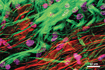
Coherent anti-Stokes Raman scattering spectroscopy, or CARS microscopy, is different. CARS microscopy images samples not by labelling, but by taking advantage of molecules intrinsic vibrational contrast, which allows for completely stain-free imaging.
CARS achieves this by using three separate femtosecond laser beams that are sent down a microscope to excite and relax defined parts of the molecule to be imaged. This process forces the molecule to release a light particle, or photon, and an image can be translated. CARS microscopy is virtually non-intrusive, and this allows for live – in vivo – samples to be imaged unharmed and in real-time video quality at a spatial resolution of about 300 nanometres.
A research group in Ottawa, Canada has been at the forefront of research into CARS microscopy since 2009 when the National Research Council Canada (NRC) and Olympus America Inc. officially opened CARSLab. “Our microscopes here can do fluorescence imaging, second harmonic imaging and CARS. With the flip of a switch, or the flip of a filter, we can image everything in one go” says Dr. Aaron Slepkov, a research associate with NRC-Olympus. “We can really build a story of what is going on inside the cell”.
Images of individual cells that make up the ear of a mouse have been achieved by CARSLab using the CARS microscopy technique. This essentially means using multiple CARS simultaneously to ‘illuminate’ the molecule of interest. ‘It blows my mind!’ says Slepkov. The CARSLab group has primarily focused on the imaging of biological systems, casting light on the intricate world of the very small with as little human interference as possible. This technique is allowing scientists to peer into cells, viruses and other living systems with as little disturbance as possible, and is paving the way for a more accurate database of knowledge concerning biological systems. The implications for disciplines such as medicine and virology are profound. The aim of a researcher is to observe and collect data while interacting with the subject of analysis as little as possible, as interaction inevitably influences the outcome of experimentation – often in ways that cannot be accounted for. The CARS technique has obvious implications for the physical sciences too. The development of microscopes and their properties generally but not exclusively, lie at the feet of physicists. College’s own Photonics Research Laboratory at CRANN, part of the School of Physics, uses and researches this technique.
CARS microscopy offers the possibility of increased imaging with the potential for interaction with the sample being dramatically reduced, and many research facilities are beginning to employ this technology as part of their daily research. This technique is advancing research into the properties of light and imaging and is allowing a closer, more accurate look into biological systems. This valuable technology is a new look into the familiar world of biological structures that could have profound consequences for future research and technologies.


